
When looking at this elegant drawing of a sleeping woman by Van Dyck, I’m immediately drawn to the shadow between her arm and torso. Guided gently by the reflected light, my eyes start to wander along the elbow—or more specifically, the olecranon process—following the contour of the tricep muscle. The olecranon is not a separate bone but rather a bony projection (process) of one of the forearm bones, hence its full name, the olecranon process. This is also where the tendon of the tricep muscle attaches. Notice the angular outline of the olecranon. Pointed shapes often indicate the presence of bones close to the skin or their projections that are not covered by muscles.
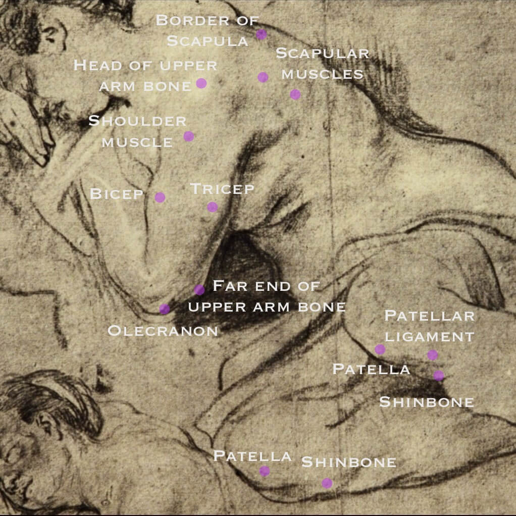
Traveling up the arm and past the armpit, you will reach the shoulder blade, also known as the scapula, which means “small shovel” in Latin. The scapula is an important attachment site for several muscles. For example, if you look closely, you can see that it is divided into two parts by a very light shading. This division reveals two scapular, or shoulder blade, muscles: the infraspinatus and teres major, but they won’t concern us at the moment.
Rather, notice how the entire upper body is bathed in light, causing some features to be lost or less visible. Van Dyck has deliberately chosen to omit some significant details, such as the collarbones, and only hint at the presence of others to reduce the anatomical information, making viewers aware of the light’s presence. Still, he has added a highlight to indicate the head of the upper arm bone pushing outward against the shoulder muscle. As a side note, the far end of the upper arm bone is also visible here—it’s the bump on the inner side of the arm above the olecranon process. The contour of the shoulder muscle is lighter compared to, for example, the biceps, emphasizing once again that this area receives direct light.
Moving on to the legs, do you recall the pointed shape of the olecranon? Now observe the numerous angular contours around the knees. These are all outlines of bones: the patella, a rounded triangular bone, also known as the kneecap, and just below it, the shinbone. The patella and shinbone are linked by the patellar ligament. Unlike tendons, which connect muscles to bones, ligaments are typically found deep within the body, connecting bones to other bones. However, the particular ligament is close to the skin surface, which makes it of interest to artists. Also, have you noticed how the calves and forearms mirror each other in shape, with their contours changing from rounded to straight? The bulging muscle bellies flatten into tendons as they approach their attachment sites around the ankles and wrists, revealing the underlying bony structure.
Lastly, let’s look at the figure as a whole. Take note of the striking twist in the sleeping woman’s torso, with her ribcage and hips turning in opposite directions while her head rests peacefully on her right arm. Her eyes are closed, yet her legs appear poised to rise. Isn’t it a perfect mix of calm and suspense, masterfully executed by Van Dyck to capture our gaze?
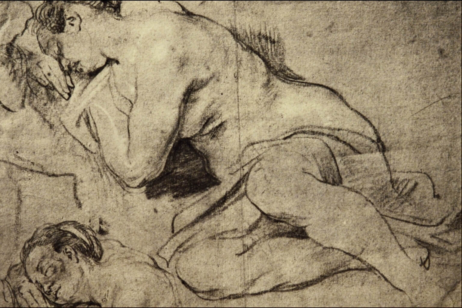
Studies of a Woman Sleeping, Anthony van Dyck
Gain invaluable insights into human anatomy that will enhance your understanding of the human form by joining our anatomy for artists life drawing workshop. Check out the schedule here.
—
About the author: Maria Kuzma-Kuzniarska, a medical illustrator and former researcher at Oxford, is the founder of Life Drawing Montmartre. For over ten years, she has been organizing bilingual life drawing workshops in Paris.